We Will Continue to be Healthy.
Speed is one of the most important factors in medical care, as it can save lives.
Digitalization makes it possible to identify the causes of illness more quickly, to make diagnoses and medical discoveries immediately, to exchange crucial information instantly and, in an emergency, to send help to where it is acutely needed without delay. We are already setting technological standards in this area. With the help of artificial intelligence and big data, diagnostic and treatment procedures are evolving rapidly. Therapies are becoming increasingly individualized and specifically tailored to each individual patient.
In the future, we will be able to detect widespread diseases such as cancer, heart attacks and strokes even earlier and treat them even more effectively.
We are already setting technological standards in this area. With the help of artificial intelligence and big data, diagnostic and treatment procedures are evolving rapidly. Therapies are becoming increasingly individualized and tailored to each patient.
For example, where doctors previously had to rely solely on the image analysis and their own personal experience to identify cancer cells on X-ray images, intelligent systems are already helping them to avoid missing any cancerous tumours. The doctor and his digital assistant both continue to learn with every single additional diagnosis, to the benefit of all future patients.
Erfolgsgeschichten

Algorithms for the Aorta!
The University of Erlangen-Nuremberg:
In her doctoral thesis, Katharina Breininger researched a method to benefit suffering from a widening of the aorta, a known aortic aneurysm. These are often treated with a minimally invasive surgery. Through the help of algorithms, the deformation of the aorta can be modelled. The big advantage is that fewer images and less contrast is necessary during the procedure. The researcher is now working on this and other topics as a professor of Artificial Intelligence in Medical Imaging at the University of Erlangen-Nuremberg.

Stay Smart throughout Pregnancy
The University of Erlangen-Nuremberg:
Record the heartbeat of the fetus at home yourself using a smartphone app or even create an ultrasound image – without an appointment at your local gynaecologist’s office or at the gynaecological clinic, saving the associated inconvenient travel and waiting times? Thanks to modern-day technology, this should soon be possible! The University of Erlangen-Nuremberg and the University Hospital Erlangen are researching the potential basis to offer the service to expectant parents.

A Clever Model With Exemplary Character
Regensburg University of Applied Sciences:
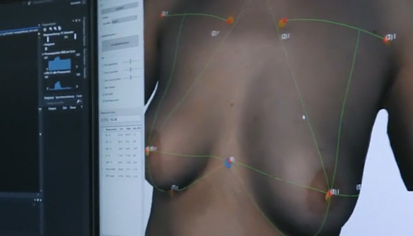
Statistical shape analysis is used in various manners in medical image processing to describe the typical shapes of organs and to be able to use them as a benchmark. In the Regensburg Medical Image Computing (ReMIC) laboratory at the Regensburg University of Applied Sciences, a statistical 3D-model of the female breast was developed from 110 breast scans. This makes it possible to generate plausible breast shapes that meet certain specifications. In the future, for example, patients will be able to have the most plausible breast shape for reconstruction after mastectomy or the most plausible result of a breast fitting with specified clinical parameters vividly displayed.
The Regensburg Breast Shape Model (RBSM) is the first openly available statistical shape model of the female breast and an important step towards the future simulation of breast surgery. It can be downloaded free of charge from the ReMIC website.
Link to the Regensburg Center for Artificial Intelligence (RCAI)
State-of-the-Art Research

Prof. Dr. Andreas M. Kist
Junior Professorship for Artificial Intelligence in Communication Disorders
The University of Erlangen-Nuremberg
Andreas Kist conducts research in the clinical application of artificial intelligence in the field of communication disorders. For example, he is working on high-speed video endoscopy of vocal folds for quantitative assessment of voice physiology and voice pathology. Simply put, imaging techniques are being used to try to put numbers on voice quality. Because our vocal folds vibrate so rapidly, several hundred times a second, images must be acquired at a correspondingly fast rate – 4,000 frames per second or more – and this volume of data must subsequently be analysed efficiently and, ideally, fully automatically. And that’s where AI comes in. The team uses artificial neural networks that are specifically adapted to process this video data so that it can then be clinically evaluated and interpreted.
(Photo: Kathrin Kist/Soulmate Photography)


